Tailored Surfaces
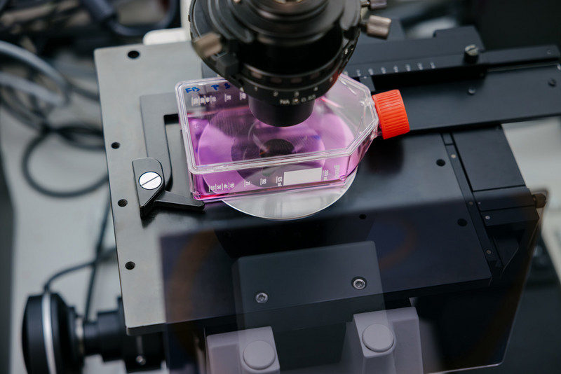
Surfaces and Nanomaterials for Targeted Interaction with Biological Systems
The IOM develops functional surfaces and nanoobjects that interact specifically with cells and tissues. The goal is to influence or control biological processes or to harness them for diagnostic and therapeutic applications.
Development of biofunctional surfaces and nanostructures
- Modification of surfaces using ion, electron, and photon technologies
- Targeted control of the interaction between materials and biological systems
- Combining experimentation and modeling to optimize functionality
- Development of materials for diagnostic and therapeutic applications
Nanostructured materials for cell interaction and tissue models
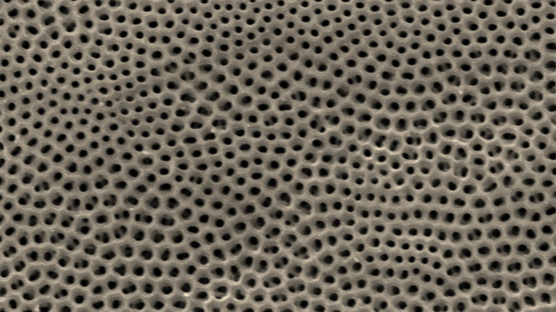
- Titanium dioxide nanotube scaffolds for long-term cultures of biological tissues
- Materials with optimized cell adhesion and surface functionality
- Use of structured surfaces to specifically influence cell behavior
- Integration of additional functions such as electrical stimulation
Magnetically active materials and nanoparticles
- Development of ferromagnetic shape-memory materials (e.g., Fe–Pd)
- Materials with superelasticity and magnetic actuability
- Synthesis of superparamagnetic iron oxide nanoparticles
- Customizable surface chemistry for biocompatible applications
Contributions to diagnostics, therapy, and theranostics
- Materials for imaging and targeted therapies
- Development of systems for theranostic applications
- Reduction of side effects through tailored material properties
- Contribution to the transfer to medical and bioanalytical applications
Publikation highlights
S. Friebe, S. Weigel, M. Francke, S. G. Mayr
Organotypic culture of adult vascularized porcine retina explants in vitro on nanotube scaffolds
Biol. Proced. Online 27 (2025) 35, https://doi.org/10.1186/s12575-025-00301-5
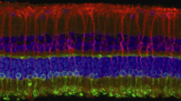
Titanium dioxide nanotubescaffolds are particularly well-suited for the long-term organotypic culture of vascularized porcine retinal explants. During culture, the morphological, histological, and biochemical integrity, as well as the vitality of the tissue, are maintained over an extended period. In the future, this will enable a powerful ex vivo model system for drug testing and diagnostics, thereby reducing the need for animal testing.
J. Dietrich, V. Wolfer, R. Konieczny, A. Lotnyk, A. Kahnt, S. G. Mayr
Electron beam-assisted synthesis of superparamagnetic iron oxide nanoparticles: reaction kinetics and structural–magnetic properties
J. Phys. Chem. C, 128 (2024) 17013-17022, https://doi.org/10.1021/acs.jpcc.4c03146
Electron beam radiolysis is used to selectively synthesize iron oxide nanoparticles with an average size of approximately 6 nm in a single-step process. The formation mechanisms are elucidated, and it is demonstrated that, as the irradiation dose increases, superparamagnetic, increasingly crystalline particles with higher saturation magnetization are formed.
A. Holm, A. Kupferer, S. Mändl, A. Lotnyk, S. G. Mayr
Conductive tracks in carbon implanted Titania nanotubes: atomic‐scale insights from experimentally based ab initio molecular dynamics modeling
Adv. Theory Simul. 5 (2022) 2200063, https://doi.org/10.1002/adts.202200063
Using ab initio molecular dynamics and density functional theory calculations, the atomic-level mechanisms underlying the structural changes in titanium dioxide nanotubes induced by carbon ion implantation are investigated. The results show that carbon chains form within the material, which—according to the calculated electronic states—specifically contribute to increased electrical conductivity.
A. Kupferer, A. Holm, A. Lotnyk, S. Mändl, S. G. Mayr
Compositional patterning in carbon implanted Titania nanotubes
Advanced Functional Materials, 31 (2021) 2104250, https://doi.org/10.1002/adfm.202104250
This paper describes the effects of low-energy ion implantation on titanium dioxide nanotubes and highlights the interplay of competing processes such as sputtering, readsorption, plastic flow, and structural and chemical rearrangement. Through experimental analyses and theoretical modeling, an intrinsically driven change in the local chemical composition is demonstrated, which leads to increased conductivity and improved material properties and opens up applications in biosensor technology and for neuroelectrodes.
S. Kallendrusch, F. Merz, I. Bechmann, S. G. Mayr, M. Zink
Long‐term tissue culture of adult brain and spleen slices on nanostructured scaffolds
Advanced healthcare materials, 6 (2017) 1601336, https://doi.org/10.1002/adhm.201601336
Biomimetic titanium dioxide nanotubescaffolds with tissue-specific properties enable long-term organotypic ex vivo culture of adult murine brain and spleen tissues. Compared to established PTFE membranes, the tissues on the nanotubes exhibit higher viability for at least two weeks, improved morphology, and reduced cell damage, making them a promising model for biological and medical research.
Equipment
- 10 MeV electron irradiation
- Ion implanter
- Environmental Scanning Electron Microscope
- Physical Properties Measurement System (PPMS)
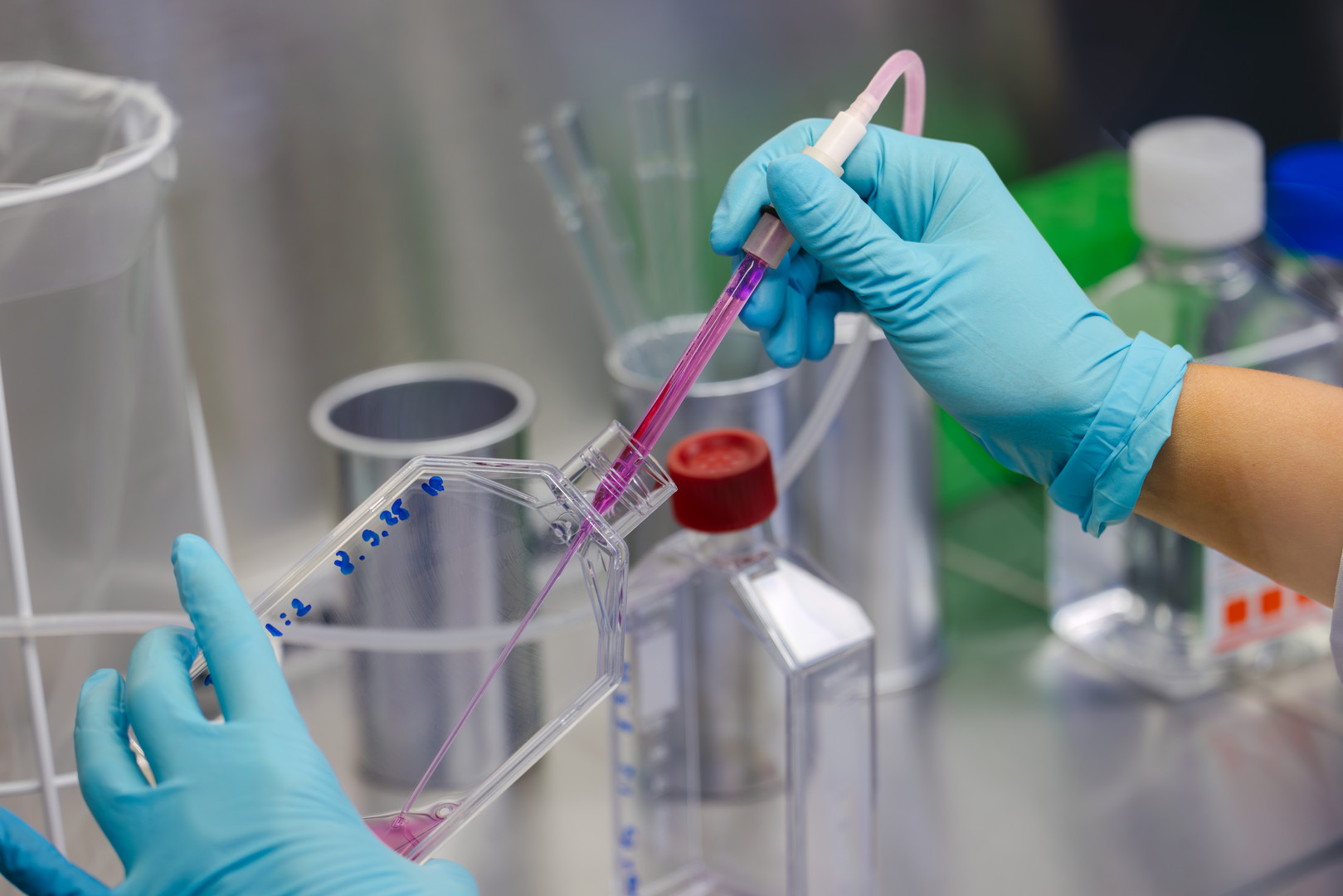
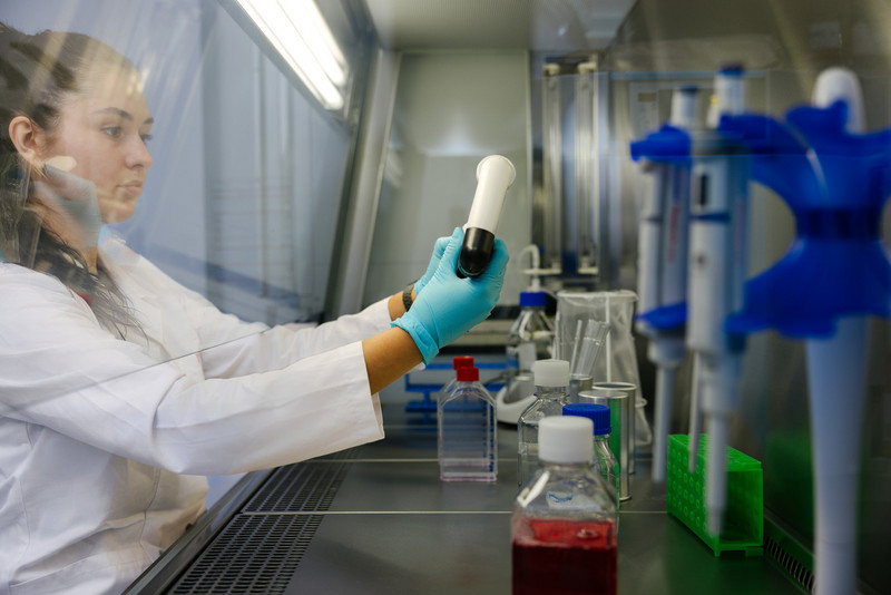
- S2 Cell and Tissue Culture Laboratory with a vibratome
- Confocal laser scanning microscope
- Ab initio modeling using density functional theory and molecular dynamics
Topics for Doctoral, Master's, and Bachelor's Theses
Akademische Qualifikationsarbeiten zu aktuellen Fragestellungen werden laufend vergeben. Bei Interesse kontaktieren Sie bitte Prof. Dr. S. G. Mayr.